lv histology cross section | normal Lv geometry lv histology cross section Overall size of the left ventricle as it appears in noncontrast CT is a composite of the ventricular volume and myocardial mass. We describe a method to estimate the LV size using a single . EBC at Night is a pool party focused nightclub that plays mostly EDM music. The nightclub is open Wednesday, Friday and Saturday in the spring and summer. The pool is only open May through September, but the club opens for business in April and often stays open into December with the pool being shut down.
0 · normal Lv shape
1 · normal Lv geometry
2 · Lv geometry diagram
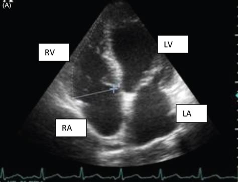
Assessment of Left Ventricular Function by Echocardiography: The Case for Routinely Adding Global Longitudinal Strain to Ejection Fraction | JACC: Cardiovascular Imaging.
normal Lv shape
a Diagrams illustrating the three cross sections of the hearts: the first mid-ventricular one and at least two additional parallel sections towards the apex. b On the left: left ventricular (posterior), . Measurements of LV mass index in patients with hypertrophy due to aortic stenosis (•) or aortic insufficiency ( ) preoperatively, in intermediate postoperative period (≈1.5 years), .The myofiber geometry of the left ventricle (LV) changes gradually from a right-handed helix in the subendocardium to a left-handed helix in the subepicardium. In this review, we associate the .
Overall size of the left ventricle as it appears in noncontrast CT is a composite of the ventricular volume and myocardial mass. We describe a method to estimate the LV size using a single .
The myofiber geometry of the left ventricle (LV) changes gradually from a right-handed helix in the subendocardium to a left-handed helix in the subepicardium. In this review, .
The histology of RV biopsy specimens obtained with a bioptome or by sharp dissection was a reliable indicator of histology in the larger RV and LV cross sections. This correlation was true . The ECG changes in a patient with left ventricular hypertrophy (LVH) were described 117 years ago by Einthoven in 1906 (Einthoven, 1957 ). He drew attention to the . Cross-sectional slices often display a macroscopic whorl-like appearance reflecting a combination of myocyte disarray and fibrosis. Mid-ventricular cross sections of human heart .
The interventricular septum separates the right and left ventricle. It is positioned obliquely, sloped to the right, and encroached onto the right ventricle. The septum bulges into .
a Diagrams illustrating the three cross sections of the hearts: the first mid-ventricular one and at least two additional parallel sections towards the apex. b On the left: left ventricular (posterior), septal (middle), and right ventricular (posterior) thickness measurements, by excluding trabeculae and papillary muscles. Measurements of LV mass index in patients with hypertrophy due to aortic stenosis (•) or aortic insufficiency ( ) preoperatively, in intermediate postoperative period (≈1.5 years), and late postoperatively (≈8 years). The LV mass index in normal adults is shown in the bar.
The myofiber geometry of the left ventricle (LV) changes gradually from a right-handed helix in the subendocardium to a left-handed helix in the subepicardium. In this review, we associate the LV myofiber architecture with emerging concepts of the electromechanical sequence in a .LV myocardial sections should include the papillary muscles. Additionally, one block from any area with significant macroscopic abnormalities should be taken. Hematoxylin and eosin and a connective tissue stain (elastic van Gieson, trichrome, or Sirius red) are routinely performed.Overall size of the left ventricle as it appears in noncontrast CT is a composite of the ventricular volume and myocardial mass. We describe a method to estimate the LV size using a single cross-section in noncontrast CT and determined normal ranges on . The myofiber geometry of the left ventricle (LV) changes gradually from a right-handed helix in the subendocardium to a left-handed helix in the subepicardium. In this review, we associate the LV myofiber architecture with emerging concepts of the electromechanical sequence in a beating heart.
The histology of RV biopsy specimens obtained with a bioptome or by sharp dissection was a reliable indicator of histology in the larger RV and LV cross sections. This correlation was true for xenografts with extensive histologic injury and for grafts with lesser degrees of rejection. The ECG changes in a patient with left ventricular hypertrophy (LVH) were described 117 years ago by Einthoven in 1906 (Einthoven, 1957 ). He drew attention to the distinctive finding—the increased QRS amplitude in the “left hand to left foot lead” (i.e., lead III).

normal Lv geometry
Cross-sectional slices often display a macroscopic whorl-like appearance reflecting a combination of myocyte disarray and fibrosis. Mid-ventricular cross sections of human heart specimens with hypertrophic cardiomyopathy were sampled in the study by . The interventricular septum separates the right and left ventricle. It is positioned obliquely, sloped to the right, and encroached onto the right ventricle. The septum bulges into the cavity of the right ventricle, and as a result, in cross-sectional images of the left ventricles, the lumen appears to be circular [20].a Diagrams illustrating the three cross sections of the hearts: the first mid-ventricular one and at least two additional parallel sections towards the apex. b On the left: left ventricular (posterior), septal (middle), and right ventricular (posterior) thickness measurements, by excluding trabeculae and papillary muscles. Measurements of LV mass index in patients with hypertrophy due to aortic stenosis (•) or aortic insufficiency ( ) preoperatively, in intermediate postoperative period (≈1.5 years), and late postoperatively (≈8 years). The LV mass index in normal adults is shown in the bar.
The myofiber geometry of the left ventricle (LV) changes gradually from a right-handed helix in the subendocardium to a left-handed helix in the subepicardium. In this review, we associate the LV myofiber architecture with emerging concepts of the electromechanical sequence in a .
LV myocardial sections should include the papillary muscles. Additionally, one block from any area with significant macroscopic abnormalities should be taken. Hematoxylin and eosin and a connective tissue stain (elastic van Gieson, trichrome, or Sirius red) are routinely performed.Overall size of the left ventricle as it appears in noncontrast CT is a composite of the ventricular volume and myocardial mass. We describe a method to estimate the LV size using a single cross-section in noncontrast CT and determined normal ranges on . The myofiber geometry of the left ventricle (LV) changes gradually from a right-handed helix in the subendocardium to a left-handed helix in the subepicardium. In this review, we associate the LV myofiber architecture with emerging concepts of the electromechanical sequence in a beating heart.The histology of RV biopsy specimens obtained with a bioptome or by sharp dissection was a reliable indicator of histology in the larger RV and LV cross sections. This correlation was true for xenografts with extensive histologic injury and for grafts with lesser degrees of rejection.
The ECG changes in a patient with left ventricular hypertrophy (LVH) were described 117 years ago by Einthoven in 1906 (Einthoven, 1957 ). He drew attention to the distinctive finding—the increased QRS amplitude in the “left hand to left foot lead” (i.e., lead III). Cross-sectional slices often display a macroscopic whorl-like appearance reflecting a combination of myocyte disarray and fibrosis. Mid-ventricular cross sections of human heart specimens with hypertrophic cardiomyopathy were sampled in the study by .

The 10 Best Budget Hotels in Las Vegas, USA. Check out our selection of great cheap hotels in Las Vegas. See the latest prices and deals by choosing your dates. Hampton Inn Las Vegas Strip South, NV 89123. South of the Las Vegas Strip, Las Vegas.
lv histology cross section|normal Lv geometry


